Electro-rheological fluids:
- Opacity, need for 3D information - rules out conventional microscopy, which gives insufficient depth discrimination. Using deconvolution methods still does not yield sufficient resolution beyond the first few layers of a colloid.
- Defects - rules out scattering techniques, which gives averaged information, while we are interested in observing real-space structures.
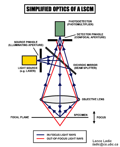
- We use laser scanning confocal microscopy, which gives good depth discrimination by rejecting most of the light from out of focus planes, as shown in the diagram below. We also use particles which are index matched to the solvent as out model ER particles, this reduces multiple scattering of light.
- Particles used - 1 micron spheres with fluorescent cores (0.2 micron)
index matched with solvent, which was a mixture of water and glycerol. The glycerol slowed the dynamics of the particles, which helped the observation of structure formation.
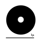
Particles synthesized by van Blaaderen,A. and Vrij,A., Langmuir 1992 ,8,2921.
Problems:
Determining structure in dense colloids
Solution:
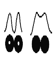
resolution ~0.6 micron in z, 0.2 micron in xy
The ER particles we use have small fluorescent cores - this enhances our ability to discriminate between individual particles. This is seen in the diagram above, where the signal from the small cores in touching particles have much less overlap than the case in which the touching particles are fluorescent throughout their volumes.